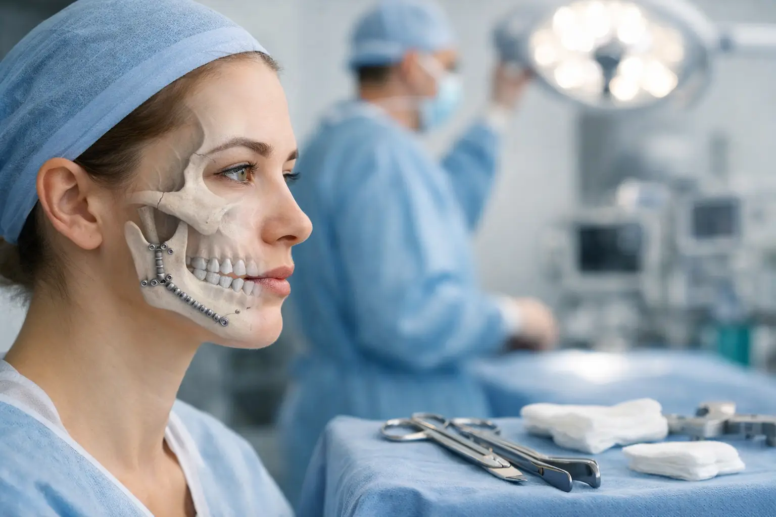
Jaw misalignment directly impacts breathing by altering airway dimensions and tongue position. When the upper jaw (maxilla) sits too far back or the lower jaw (mandible) is recessed, the airway space behind the tongue narrows. This structural relationship explains why many patients seeking orthognathic surgery report both aesthetic concerns and breathing difficulties.
The connection becomes clearer when examining airway anatomy. Your jaw bones form the structural foundation for soft tissues, including the tongue, soft palate, and pharyngeal walls. Abnormal jaw positioning forces these tissues backward, reducing the space available for airflow. During sleep, when muscle tone naturally decreases, this narrowing worsens significantly.
Jaw Misalignments That Affect Breathing
Retrognathic Mandible (Recessed Lower Jaw)
A recessed lower jaw pushes the tongue base posteriorly, directly encroaching on the airway space. This condition manifests as a weak chin profile and often accompanies Class II malocclusion, where upper teeth significantly overlap lower teeth. The tongue, anchored to the mandible, follows the jaw’s backward position and crowds the oropharyngeal airway.
Patients with retrognathic mandibles frequently develop compensatory head postures, tilting their heads back to open the airway. This adaptation leads to neck strain and temporomandibular joint stress over time. Sleep studies in these patients often reveal increased respiratory effort and fragmented sleep patterns even without frank obstructive sleep apnea.
Maxillary Hypoplasia (Underdeveloped Upper Jaw)
Underdevelopment of the maxilla creates both nasal and oral breathing challenges. The maxilla forms the floor of the nasal cavity, so inadequate growth restricts nasal passage dimensions. Patients present with a sunken midface appearance and often report chronic nasal congestion despite the absence of allergic or inflammatory conditions.
The narrow maxilla forces the tongue into a higher, more posterior position to fit within the reduced oral cavity space. This tongue positioning further compromises the airway at the velopharyngeal and oropharyngeal levels. Many patients unconsciously adopt mouth breathing patterns, leading to dry mouth, increased dental decay risk, and altered facial growth patterns in younger patients.
Vertical Maxillary Excess (Long Face Syndrome)
Excessive vertical growth of the maxilla creates an elongated lower face and an incompetent lip seal. The increased facial height prevents comfortable lip closure, forcing habitual mouth breathing. The mandible rotates downward and backward to accommodate the vertical excess, simultaneously reducing airway dimensions.
These patients exhibit specific clinical signs: increased lower facial height, gummy smile, and mentalis muscle strain when attempting lip closure. The open mouth posture allows the tongue to drop from its normal palatal position, reducing palatal width over time and creating a self-perpetuating cycle of airway compromise.
How Orthognathic Surgery Improves Breathing
Mandibular Advancement
Advancing the mandible through bilateral sagittal split osteotomy directly increases airway dimensions. The procedure involves precise cuts through the mandibular ramus, allowing forward repositioning of the entire lower jaw. Typical advancements range from 5-12mm, with each millimeter of advancement correlating to measurable airway volume increases.
Post-surgical imaging consistently demonstrates enlarged retroglossal and retropalatal airway spaces. The genioglossus muscle, attached to the inner mandible, pulls the tongue base forward with jaw advancement. This mechanical coupling explains the dramatic breathing improvements many patients experience immediately post-surgery, even before swelling fully resolves.
Maxillary Advancement and Expansion
Le Fort I osteotomy allows three-dimensional repositioning of the entire maxilla. Forward movement increases nasal cavity depth while simultaneously providing more tongue space. When combined with surgical expansion, the procedure widens both nasal passages and the oral cavity. Modern techniques achieve 5-7mm of transverse expansion through precise bone cuts and controlled distraction.
The breathing improvements from maxillary procedures extend beyond simple volume increases. Advancing the maxilla improves the nasal valve angle, the narrowest part of the nasal airway. Widening the piriform aperture and nasal floor creates laminar airflow patterns, reducing the work of nasal breathing.
Genioplasty (Chin Surgery)
While primarily considered an aesthetic procedure, genioplasty significantly impacts breathing when combined with jaw surgery. Advancing the chin bone (genial tubercles) pulls forward the genioglossus and geniohyoid muscles. This anterior traction maintains tongue position and prevents posterior collapse during sleep.
Functional genioplasty differs from simple cosmetic augmentation by focusing on muscle attachment preservation and advancement. Typical advancements of 6-10mm provide both aesthetic balance and functional airway support. The procedure adds minimal surgical time when performed with orthognathic surgery.
Breathing Improvements After Surgery
Immediate Changes
Patients often notice breathing improvements within days of surgery, despite post-operative swelling. The mechanical repositioning of jaw structures immediately enlarges the airway, though soft tissue edema temporarily masks the full benefit. Nasal breathing typically improves first as maxillary procedures correct septal deviations and turbinate hypertrophy through improved anatomical relationships.
Sleep partners frequently report eliminated or reduced snoring even during the early recovery period. This immediate change occurs because the structural airway enlargement overcomes the temporary inflammatory narrowing from surgical trauma. Patients describe the sensation as “breathing through a larger tube” once initial congestion clears.
Long-term Benefits
Six months post-surgery, when bone healing completes and soft tissues fully adapt, maximum breathing improvements manifest. Polysomnography studies at this time point show reduced respiratory disturbance indices, increased oxygen saturation nadirs, and improved sleep architecture. Many patients previously dependent on CPAP therapy achieve successful device weaning under medical supervision.
The breathing improvements remain stable long-term due to the permanent skeletal changes. Unlike soft tissue procedures that may relapse, bony advancement maintains airway patency indefinitely. Ten-year follow-up studies demonstrate sustained improvements in both subjective breathing quality and objective airway measurements.
💡 Did You Know?
Jaw surgery planning now incorporates computational fluid dynamics modeling to predict post-surgical airflow patterns. This technology allows surgeons to optimize jaw positioning for both aesthetic and functional breathing outcomes.
Diagnostic Process
Clinical Evaluation
Comprehensive breathing assessment begins with detailed history taking. Surgeons evaluate daytime fatigue, morning headaches, witnessed apneas, and positional breathing preferences. Physical examination includes nasopharyngoscopy to visualize the entire upper airway and identify specific obstruction sites.
Mallampati scoring, neck circumference measurement, and lateral profile analysis provide additional clinical data. Surgeons perform Müller’s maneuver – having patients inspire against a closed airway while visualizing collapse patterns through flexible endoscopy. This dynamic assessment reveals which airway segments will benefit most from skeletal advancement.
Imaging Studies
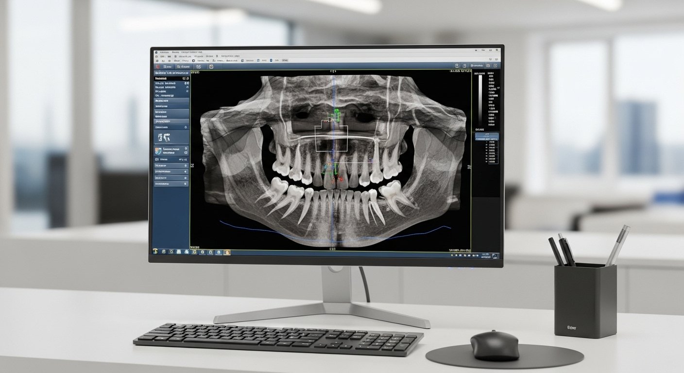
Cone beam computed tomography (CBCT) provides three-dimensional airway analysis with minimal radiation exposure. Software segmentation creates accurate volumetric measurements at defined anatomical levels. Surgeons evaluate minimum cross-sectional areas, typically located behind the soft palate or tongue base, as these predict airway resistance.
Lateral cephalometric radiographs, while two-dimensional, offer valuable measurements including posterior airway space and hyoid bone position. The relationship between these measurements and planned surgical movements guides treatment planning. Modern planning software simulates post-surgical airway changes based on proposed jaw movements.
Sleep Studies
Formal polysomnography remains the gold standard for diagnosing sleep-disordered breathing. The study records multiple physiologic parameters, including airflow, respiratory effort, oxygen saturation, and sleep stages. An apnea-hypopnea index (AHI) above 5 events per hour indicates sleep apnea, with severity classifications guiding surgical planning.
Home sleep tests provide screening alternatives for straightforward cases. These portable monitors record respiratory parameters in the patient’s normal sleep environment. While less comprehensive than laboratory polysomnography, they effectively identify moderate to severe sleep apnea requiring treatment.
What Our Orthognathic Surgeon Says
“Breathing improvement often motivates patients to pursue jaw surgery even more than aesthetic concerns. The correlation between jaw position and airway dimension is remarkably consistent – we can predict with high accuracy how much breathing will improve based on planned jaw movements.
Modern 3D planning allows us to optimize both facial aesthetics and airway function simultaneously. I’ve seen patients transform from CPAP-dependent to normal breathing after appropriate surgical correction.
Many patients don’t realize their chronic fatigue, morning headaches, and concentration difficulties stem from subtle nighttime breathing issues related to jaw position. When we correct the underlying skeletal problem, these symptoms often resolve completely.”
Treatment Planning Considerations
Combined Procedures
Optimal breathing outcomes often require addressing multiple anatomical levels simultaneously. Maxillomandibular advancement (MMA) combines upper and lower jaw advancement, providing significant airway enlargement. Typical combined advancements of 8-10mm achieve AHI reductions exceeding those of isolated procedures.
Surgeons may incorporate adjunctive procedures based on individual anatomy. Turbinate reduction improves nasal airflow when hypertrophy exists. Septoplasty corrects deviated septums that would otherwise limit breathing improvements from maxillary advancement. These combined approaches address breathing comprehensively rather than focusing on single obstruction sites.
Surgical Sequencing
Single-jaw procedures may suffice for specific breathing issues. Isolated mandibular advancement effectively treats tongue-base obstruction when the maxilla position is adequate. Conversely, isolated maxillary advancement with expansion addresses nasal obstruction and narrow palates without mandibular involvement.
The surgical team considers stability, healing patterns, and patient goals when sequencing procedures. Some patients benefit from staged surgeries, allowing soft tissue adaptation between procedures. Others achieve optimal results through single-stage comprehensive correction.
⚠️ Important Note
Not all breathing issues require jaw surgery. Mild sleep-disordered breathing may respond to conservative treatments including oral appliances, positional therapy, or continuous positive airway pressure (CPAP). Surgical intervention is typically reserved for anatomical abnormalities or failed conservative management.
Recovery and Breathing Adaptation
Early Recovery Phase
The first two weeks post-surgery involve expected swelling and congestion that temporarily affects breathing. Surgeons prescribe decongestants and saline rinses to maintain nasal patency during healing. Head elevation during sleep reduces edema and improves breathing comfort. Most patients transition from mouth to nasal breathing as swelling subsides around week three.
Elastic guidance during orthodontic finishing maintains the new jaw position while muscles adapt. This orthodontic phase, lasting 6-12 months, allows fine-tuning of the occlusion without compromising achieved airway improvements. Patients report progressive breathing improvements throughout this adaptation period.
Breathing Exercises
Post-surgical breathing exercises accelerate functional recovery. Diaphragmatic breathing techniques reduce accessory muscle use and establish efficient breathing patterns. Surgeons recommend specific exercises including alternate nostril breathing to ensure balanced nasal airflow and pursed-lip breathing to maintain positive airway pressure.
Speech therapy may benefit select patients, particularly those with long-standing compensatory tongue positions. Therapists teach proper tongue posture and swallowing patterns that support the new airway dimensions. These exercises prevent the tongue from reverting to old positions that could compromise breathing improvements.
Putting This Into Practice
- Schedule a comprehensive evaluation if you experience both jaw misalignment and breathing difficulties – the conditions are often related
- Document your breathing symptoms, including snoring patterns, daytime fatigue levels, and sleep quality, before consultation
- Obtain any previous sleep studies or imaging that may help in treatment planning
- Consider an orthodontic consultation, as most jaw surgeries require coordinated orthodontic treatment
- Discuss all breathing concerns during surgical planning, as positioning can be optimized for both aesthetic and functional outcomes
When to Seek Professional Help
- Witnessed pauses in breathing during sleep
- Chronic daytime fatigue despite adequate sleep hours
- Morning headaches occurring more than twice weekly
- Difficulty breathing through your nose, requiring constant mouth breathing
- Jaw appearance concerns combined with any breathing symptoms
- Previous CPAP intolerance with documented sleep apnea
- Dry mouth upon waking, suggesting nighttime mouth breathing
- Partner complaints about loud snoring affecting their sleep
Commonly Asked Questions
How long after jaw surgery will my breathing improve?
Initial improvements occur within 1-2 weeks as surgical swelling decreases. Significant improvements manifest by 6-8 weeks, with maximum benefit achieved at 6 months when all tissues have fully healed and adapted to the new jaw position.
Can jaw surgery eliminate my need for CPAP?
Many patients with anatomically caused sleep apnea successfully discontinue CPAP after appropriate jaw advancement. Success depends on the severity of pre-surgical AHI and the amount of advancement achieved. Post-surgical sleep studies at 6 months determine CPAP necessity.
Will insurance cover jaw surgery if I have sleep apnea?
Coverage varies significantly between insurance plans. Documented sleep apnea with failed CPAP therapy strengthens medical necessity claims. Pre-authorization typically requires sleep study results, CPAP compliance data, and comprehensive clinical documentation.
What’s the difference between jaw surgery and other sleep apnea surgeries?
Jaw surgery addresses the skeletal framework causing airway obstruction, while soft tissue procedures like UPPP remove or reposition tissues. Skeletal advancement provides more predictable, stable results for appropriately selected patients with jaw-related airway obstruction.
How do surgeons determine the right amount of advancement?
Surgeons use 3D imaging analysis, airway measurements, and aesthetic principles to plan movements. Virtual surgical planning allows visualization of post-surgical results before surgery. The goal balances maximum airway improvement with facial harmony and stable dental occlusion.
Next Steps
Jaw surgery offers permanent solutions for breathing issues caused by skeletal misalignment. The procedures simultaneously improve facial aesthetics and airway function through precise repositioning of the facial skeleton.
If you’re experiencing breathing difficulties along with jaw misalignment, our MOH-accredited oral and maxillofacial surgeons provide comprehensive evaluation and personalized treatment planning for orthognathic surgery.