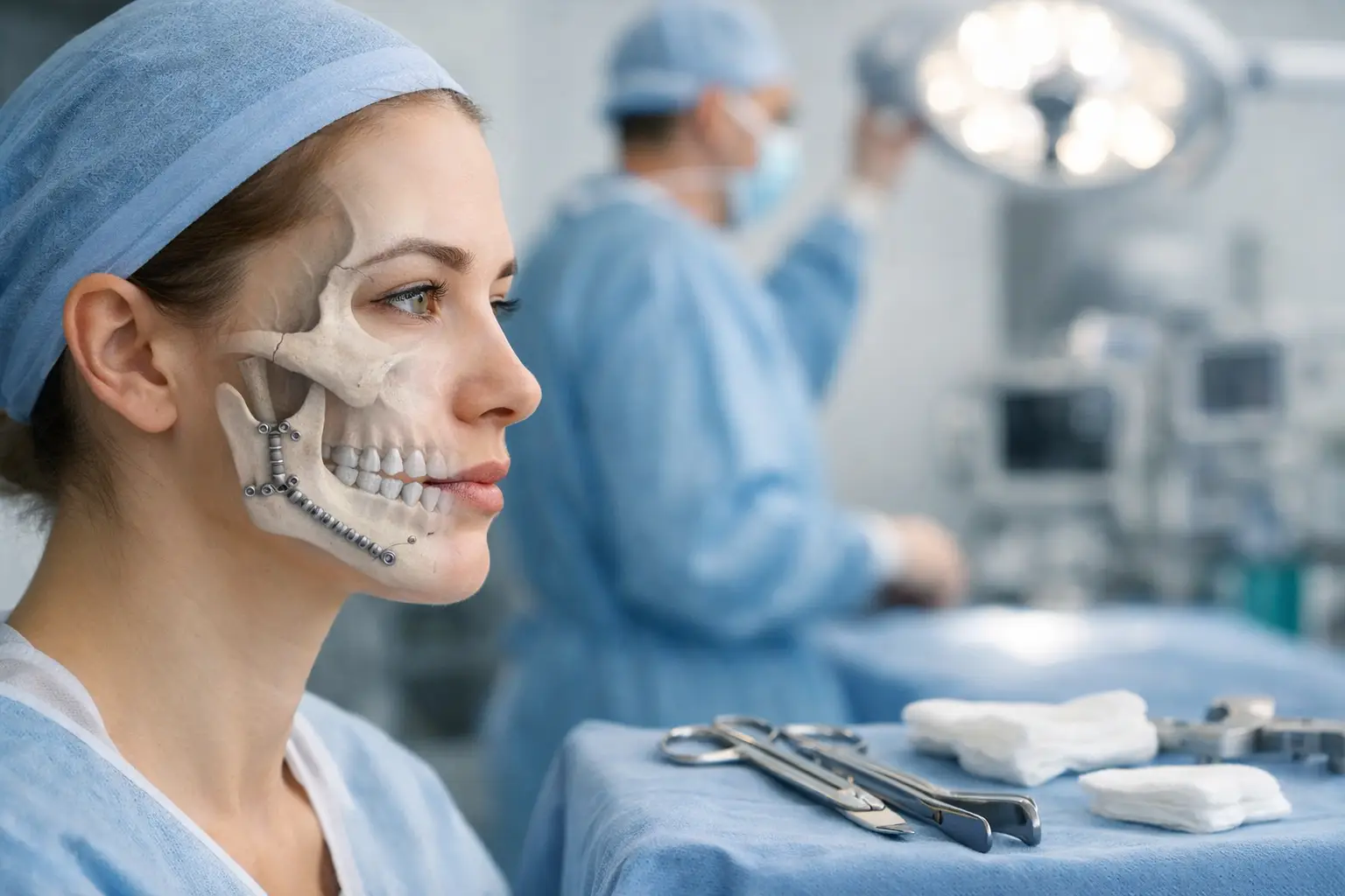
3D imaging has transformed oral surgery, moving from two-dimensional X-rays to visualising the entire maxillofacial complex in 3D. Cone Beam Computed Tomography (CBCT) produces detailed volumetric images of bone density, nerve pathways, and anatomical structures with a resolution of 0.076-0.4mm. This technology enables surgeons to precisely measure bone thickness, identify the exact path of the inferior alveolar nerve, and plan implant placement with submillimeter accuracy. Unlike traditional panoramic X-rays, which compress 3D structures into 2D images, CBCT captures data from multiple angles to digitally reconstruct the jaw, teeth, and surrounding tissues. The resulting images allow rotation, cross-sectioning, and measurement in any plane, providing comprehensive visualisation for complex procedures.
CBCT Technology in Oral Surgery
CBCT scanners use a cone-shaped X-ray beam that rotates 180-360 degrees around the patient’s head, capturing hundreds of projection images in 10-40 seconds. The computer reconstructs these projections into a three-dimensional dataset with voxel sizes ranging from 0.076mm to 0.4mm. This resolution reveals cortical bone boundaries, trabecular patterns, and the periodontal ligament space.
The technology produces three primary image types: axial slices showing horizontal cross-sections, coronal views displaying front-to-back sections, and sagittal images revealing side views. Surgeons can generate panoramic reconstructions, create 3D surface renderings, and extract specific regions of interest. CBCT’s focused field-of-view options, ranging from 5cm for single teeth to 23cm for full craniofacial imaging, optimise radiation dose while capturing necessary anatomy.
Radiation exposure from CBCT ranges from 19 to 674 microsieverts, depending on scanner settings and field size, compared to 2.7 to 24 microsieverts for panoramic radiographs. Modern CBCT units incorporate dose-reduction protocols, including collimation, pulsed exposure, and automatic exposure control, to minimise patient radiation exposure while maintaining diagnostic quality.
Surgical Planning Applications
Virtual surgical planning transforms CBCT data into interactive 3D models, allowing surgeons to simulate procedures before entering the operating room. Software platforms allow measurement of bone dimensions, calculation of angulations, and virtual osteotomy planning with 0.5mm precision. In orthognathic surgery, surgeons can predict post-operative facial changes by digitally repositioning jaw segments and visualising soft-tissue responses.
Implant planning software overlays CBCT data with digital impressions to design optimal implant positions considering available bone volume, prosthetic requirements, and anatomical limitations. The software calculates bone density in Hounsfield units, identifies the mandibular canal’s position within 1mm accuracy, and determines safe implant lengths. Surgeons can virtually place implants, assess primary stability based on bone quality, and design custom surgical guides.
In reconstructive procedures, 3D imaging enables precise measurement of defect volumes, assessment of donor sites, and virtual bone-graft design. Mirror imaging techniques use the unaffected side as a template for reconstruction, while segmentation tools isolate specific anatomical structures for detailed analysis. Pre-bent reconstruction plates can be manufactured based on 3D models, reducing operative time by 30-60 minutes.
Navigation and Guidance Systems
Computer-assisted navigation systems merge pre-operative CBCT data with real-time surgical positioning, achieving accuracy within 1-2mm. Optical tracking systems use infrared cameras to monitor reflective markers attached to surgical instruments and the patient, displaying instrument position on the 3D model throughout surgery. This technology is particularly valuable near structures such as the inferior alveolar nerve or the maxillary sinus.
Dynamic navigation enables real-time drill tracking during implant placement, displaying depth, angulation, and proximity to structures on-screen. The system provides visual and auditory warnings when approaching predetermined safety margins, typically set at 2mm from anatomy. Studies demonstrate that navigation-guided implant placement achieves angular deviations of less than 4 degrees and apical deviations of less than 1.5mm from planned positions.
Static guidance using 3D-printed surgical templates offers an alternative approach, with tooth-supported guides achieving placement accuracy within 1.2mm at the implant apex and 5 ° angular deviation. Bone-supported guides used in fully edentulous cases demonstrate similar accuracy when properly stabilised with fixation pins.
Complex Case Management
3D imaging reveals pathology invisible on conventional radiographs, including small periapical lesions, root fractures, and early-stage osteonecrosis. CBCT identifies variations in root canal anatomy, accessory canals, and the relationships between roots and adjacent structures. For impacted teeth, 3D imaging precisely locates tooth position, root morphology, and proximity to nerves or sinuses, informing surgical approach decisions.
Trauma cases benefit from 3D visualisation of fracture patterns, fragment displacement, and associated soft tissue injuries. CBCT with a resolution of 0.2mm or better can detect non-displaced fractures missed on plain radiographs. The technology enables accurate measurement of fracture gaps, assessment of bony union during healing, and evaluation of the position of post-surgical hardware.
Pathological lesions appear three-dimensionally, allowing volumetric measurement, margin assessment, and surgical approach planning. Surgeons can determine cortical perforation, measure distances to structures, and plan resection margins. For cystic lesions, 3D imaging differentiates between single and multilocular patterns, guiding treatment decisions between enucleation and decompression.
💡 Did You Know?
Modern CBCT software can automatically detect and highlight the inferior alveolar nerve canal using artificial intelligence algorithms, reducing nerve identification time from minutes to seconds while maintaining accuracy comparable to manual tracing by radiologists.
Patient Communication Benefits
3D visualisations transform patient consultations by displaying anatomy and pathology in intuitive formats. Patients who view their own 3D reconstructions demonstrate a better understanding of proposed procedures than those who receive traditional X-ray explanations. Color-coded models highlight different structures—bone in white, nerves in yellow, teeth in natural colors—making complex anatomy accessible.
Treatment simulations show patients’ expected outcomes before surgery, including facial profile changes in orthognathic cases or final implant positions in reconstructive procedures. Virtual before-and-after comparisons help set realistic expectations and facilitate informed consent discussions. Interactive models allow patients to rotate and explore their anatomy and ask specific questions about their condition.
Surgical guides and models produced from 3D data serve as tangible consultation tools. Patients can handle 3D-printed models of their jaw to understand the defect size or planned reconstruction. This tactile experience, combined with visual displays, improves patient confidence in treatment plans.
What Our Oral Surgeon Says
3D imaging fundamentally changes how we approach complex cases. When I can visualise the exact relationship between an impacted tooth and the inferior alveolar nerve before surgery, I can plan the most conservative approach that preserves sensation. The ability to measure available bone volume in three dimensions means we can often avoid bone grafting procedures that seemed necessary based on 2D imaging alone. Showing patients their actual anatomy on screen transforms abstract explanations into a concrete understanding, leading to better treatment acceptance and compliance.
Future Developments
Artificial intelligence integration promises automated pathology detection, with algorithms currently achieving accuracy comparable to that of specialist radiologists for specific conditions. Machine learning models trained on thousands of CBCT scans can identify periapical lesions, automatically measure them, and flag abnormalities for clinical review.
Augmented reality systems under development overlay 3D imaging data directly onto the surgical field through specialised glasses or microscope displays. Early clinical trials demonstrate the feasibility of guided osteotomies and implant placement, though widespread adoption awaits hardware refinement and cost reduction.
Integration with intraoral scanners creates complete digital workflows from diagnosis through restoration. Combined datasets allow virtual articulation, occlusal analysis, and prosthetic design based on actual jaw relationships captured in CBCT. Real-time CBCT imaging during surgery remains experimental but could enable immediate verification of surgical outcomes.
⚠️ Important Note
While 3D imaging provides exceptional detail, interpretation requires specialized training. Incidental findings occur frequently in CBCT scans, requiring careful evaluation to distinguish clinically significant pathology from normal anatomical variations or artifacts.
Putting This Into Practice
- Request CBCT imaging when conventional X-rays leave diagnostic uncertainty about tooth positions, bone availability, or the extent of pathology.
- Ask to view your 3D images during the consultation to better understand your specific anatomy and planned procedures.
- Inquire about surgical guide options for implant procedures, particularly in aesthetic zones or near structures.
- Discuss whether navigation technology could benefit your specific case, especially for procedures near nerves or sinuses.
- Request printed 3D models for complex reconstructive cases to visualise the planned outcome.
When to Seek Professional Help
- Persistent jaw pain with normal-appearing conventional X-rays
- Failed previous oral surgery with unexplained complications
- Multiple missing teeth requiring comprehensive reconstruction
- Facial asymmetry or bite problems affecting function
- Impacted wisdom teeth with symptoms
- Jaw cysts or tumours requiring surgical removal
- Facial trauma with suspected fractures
- Planning for dental implants in areas with limited bone
Commonly Asked Questions
How does 3D imaging radiation compare to regular dental X-rays?
CBCT radiation varies with scan size and settings, typically delivering 19-674 microsieverts, compared with 2.7-24 microsieverts for panoramic X-rays. Small-field CBCT scans focused on specific areas use radiation doses comparable to 2-3 panoramic radiographs, whereas full-skull scans require higher doses, justified only for complex surgical planning.
Will insurance cover 3D imaging for my oral surgery?
Coverage varies by provider and procedure. Medical necessity documentation typically includes surgical complexity, proximity to structures, or previous surgical complications. Pre-authorisation often requires the submission of conventional radiographs that demonstrate limitations for surgical planning.
How long before surgery should 3D imaging be performed?
CBCT scans remain diagnostically accurate for 3-6 months in stable conditions. Trauma cases require imaging within days of injury, while elective procedures can use scans up to 6 months old if no interim dental work occurs. Progressive pathology mandates imaging closer to the surgical date.
Can 3D imaging replace all traditional dental X-rays?
CBCT excels at surgical planning but doesn’t replace all conventional imaging. Intraoral radiographs provide better resolution for detecting cavities and assessing periodontal bone levels. CBCT serves specific diagnostic purposes when 3D visualisation adds clinical value beyond that of 2D imaging.
What’s the difference between medical CT and dental CBCT?
Dental CBCT uses cone-shaped beams optimised for hard-tissue imaging, with lower radiation doses than medical CT. CBCT provides bone detail with a resolution of 0.076-0.4mm but limited soft-tissue contrast. Medical CT offers better soft-tissue visualisation but requires higher radiation exposure and typically lower hard-tissue resolution.
Next Steps
3D imaging transforms oral surgery from estimation to precision, enabling accurate planning and predictable outcomes. Virtual surgical planning reduces operative time while navigation systems enhance safety near structures.
If you’re experiencing complex dental problems, facial trauma, or considering reconstructive procedures, our Oral Surgeon specialises in 3D imaging techniques for comprehensive evaluation and treatment planning.