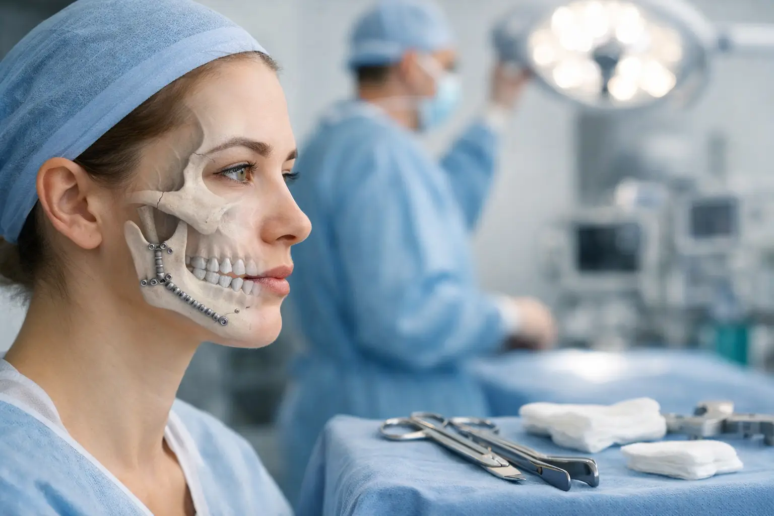
Facial trauma affecting the jaw requires specialised surgical intervention to restore both function and appearance. Whether from motor vehicle accidents, falls, sports injuries, or physical assault, jaw fractures and dislocations demand prompt medical attention and often complex surgical repair. The path from injury to recovery involves multiple stages, each requiring specific care approaches and realistic expectations.
Jaw surgery after facial trauma encompasses various procedures depending on injury severity and location. Mandibular fractures, maxillary injuries, and temporomandibular joint damage each present unique surgical challenges. Modern reconstructive techniques allow surgeons to address both immediate structural damage and long-term functional concerns, though the journey requires patience and adherence to post-operative protocols.
Types of Jaw Injuries Requiring Surgery
Mandibular Fractures
The mandible (lower jaw) absorbs significant force during facial trauma, making it particularly vulnerable to fractures. Common fracture sites include the body, the angle, the condyle, and the symphysis. Each location presents distinct surgical challenges and recovery considerations.
Condylar fractures affect the jaw joint directly, potentially causing long-term bite problems if not correctly aligned. Body fractures often result in visible facial asymmetry and immediate difficulty opening the mouth. Angle fractures frequently occur at the wisdom tooth region, where the bone naturally thins. Symphysis fractures at the centre of the chin can affect both sides of the jaw simultaneously.
Maxillary Injuries
Upper jaw fractures follow specific patterns classified as Le Fort I, II, or III injuries. Le Fort I fractures run horizontally above the teeth, separating the tooth-bearing portion from the upper face. Le Fort II fractures create a pyramid-shaped break extending through the nose and cheekbones. Le Fort III fractures completely separate the facial bones from the skull base.
Each pattern requires different surgical approaches and fixation methods. Higher-level fractures often involve multiple facial structures, necessitating coordination between jaw surgeons and other specialists.
Temporomandibular Joint Damage
TMJ injuries range from simple dislocations to complex fractures involving the joint surfaces. Disc displacement, capsular tears, and articular surface damage can result in chronic pain and limited jaw movement without proper treatment.
Joint injuries often accompany other facial fractures, complicating the surgical planning process. Some cases require arthroscopic intervention or open joint surgery to restore normal function.
Surgical Planning and Preparation
Initial Assessment
Comprehensive imaging underpins surgical planning. CT scans provide three-dimensional views of bone injuries, while MRI scans reveal soft-tissue damage. Dental impressions and bite registrations document pre-injury occlusion when possible.
Surgeons assess airway patency, neurological function, and associated injuries during the initial evaluation. Damage to facial nerves, tear ducts, or salivary glands influences surgical timing and approach. Some patients require immediate airway management before definitive jaw repair.
Pre-operative Considerations
Medical optimisation addresses underlying conditions that could affect healing. Nutritional status, smoking cessation, and diabetes control directly impact surgical outcomes. Patients taking blood thinners require medication adjustments before surgery.
A dental evaluation identifies damaged teeth that require extraction or repair. Existing dental work may need modification to accommodate surgical hardware. Poor oral hygiene increases the risk of infection, making preoperative dental cleaning important in stable patients.
Timing of Surgery
Immediate surgery within 24-48 hours offers advantages for simple, isolated fractures. Fresh fracture edges align more easily, and soft tissue swelling remains minimal. However, severe swelling, associated injuries, or medical instability may necessitate delayed intervention.
Delayed surgery after 7-14 days allows swelling resolution and medical stabilisation but makes fracture reduction more challenging. Scar tissue formation and early bone healing complicate anatomical restoration. Surgeons balance these factors when determining optimal timing for each case.
Surgical Procedures
Open Reduction and Internal Fixation
ORIF remains the standard for most displaced jaw fractures. Surgeons access fracture sites through carefully planned incisions, either inside the mouth or through small external approaches. Direct visualisation enables precise anatomical reduction before fixation hardware is applied.
Titanium plates and screws provide stable fixation while bones heal. Plate design varies by fracture location and pattern. Miniplates suffice for simple fractures, while reconstruction plates span larger defects. Biodegradable fixation offers advantages in specific situations, particularly in growing patients.
Closed Reduction Techniques
Non-displaced or minimally displaced fractures may heal with closed reduction and immobilisation. Intermaxillary fixation (IMF) using arch bars, wires, or screws maintains proper bite relationships during healing. This approach avoids surgical incisions but requires 4-6 weeks of jaw immobilisation.
Modern IMF techniques minimise patient discomfort while maintaining stability. Bone-supported devices eliminate the need for circumdental wires, reducing gingival trauma. Some systems allow limited jaw movement for improved nutrition and oral hygiene.
Bone Grafting
Comminuted fractures, bone loss, or delayed treatment may necessitate bone grafting. Autogenous bone from the hip, rib, or skull provides optimal healing potential. Processed allograft or synthetic materials offer alternatives when donor site morbidity concerns exist.
Grafting techniques vary by defect size and location. Particulate grafts fill small gaps, while structural grafts bridge larger defects. Guided bone regeneration membranes prevent soft tissue ingrowth during healing.
Recovery Timeline
Immediate Post-operative Period (Days 1-7)
The first week focuses on pain control, swelling management, and infection prevention. Facial swelling peaks around day 3-4, then gradually subsides. Ice application, head elevation, and prescribed medications minimise discomfort.
Dietary restrictions begin immediately, with liquid nutrition important for IMF patients—those without jaw wiring progress to soft, non-chew foods. Meticulous oral hygiene with prescribed rinses and specialised tools helps prevent infection around surgical sites.
Early Recovery Phase (Weeks 2-6)
Suture removal occurs at 7-10 days for external incisions. Intraoral sutures often dissolve spontaneously. Regular follow-up appointments monitor healing progress and occlusion stability.
Physical therapy may begin for TMJ injuries or prolonged immobilisation cases. Gentle range-of-motion exercises prevent permanent stiffness. Patients with IMF experience gradual elastic guidance rather than rigid fixation during this period.
Bone Healing Phase (Weeks 6-12)
Radiographic evidence of bone healing appears around 6-8 weeks. IMF removal allows progressive return of jaw function. Initial jaw movements feel stiff and uncoordinated, but they improve with regular exercise.
Dietary advancement proceeds cautiously, avoiding hard or chewy foods that stress healing bones. Patients often require nutritional counselling to regain weight lost during the liquid diet phase.
Long-term Recovery (Months 3-12)
Complete bone remodelling continues for several months. Facial sensation returns gradually if nerve involvement occurred. Some patients experience weather-related discomfort around surgical sites during the first year.
Hardware removal may be necessary for symptomatic plates or at the patient’s preference. This minor procedure typically occurs after 6-12 months when bone healing is complete.
Potential Complications
Infection
Surgical site infections occur more frequently with intraoral approaches and contaminated wounds. Signs include increasing pain, swelling, drainage, and fever. Early recognition and appropriate antibiotics usually resolve infections without hardware removal.
Osteomyelitis represents a serious complication requiring prolonged antibiotic therapy and possible surgical debridement. Risk factors include poor oral hygiene, smoking, and immunocompromise.
Malocclusion
Improper fracture reduction or inadequate fixation results in bite abnormalities. Patients usually report premature tooth contact, jaw deviation, or an inability to bring the teeth together. Minor discrepancies may resolve with orthodontic treatment, while major malunions require revision surgery.
Non-union or Delayed Union
Fractures that fail to heal within expected timeframes require investigation into underlying causes. Infection, inadequate fixation, or poor bone quality contribute to non-union. Treatment involves revision surgery with bone grafting and rigid fixation.
Nerve Injury
Inferior alveolar nerve damage causes lower lip and chin numbness. Mental nerve injury specifically affects lip sensation. Most nerve injuries from trauma or surgery recover within 6-12 months, though permanent deficits occasionally persist.
TMJ Dysfunction
Post-traumatic arthritis, disc displacement, or ankylosis can develop months to years after injury. Symptoms include pain, clicking, locking, or progressive limitation of mouth opening. Management ranges from conservative therapy to joint replacement in severe cases.
Rehabilitation and Physical Therapy
Early Mobilization
Controlled jaw exercises begin immediately after IMF removal or when surgically appropriate. Initial movements focus on gentle opening and closing within pain-free ranges. Lateral movements and protrusion exercises follow as comfort improves.
Manual therapy by trained physiotherapists addresses muscle spasms and joint restrictions. Techniques include gentle stretching, joint mobilisation, and soft tissue massage. Home exercise programs reinforce clinical treatments.
Strengthening Exercises
Progressive resistance exercises rebuild jaw muscles weakened by disuse. Patients use rubber tubing, tongue depressors, or specialised devices for controlled strengthening. Proper technique prevents excessive joint stress during the rehabilitation phase.
Functional exercises simulate normal jaw activities like chewing and speaking. Graduated food textures challenge the recovering jaw while building confidence. Speech therapy addresses articulation problems from altered jaw relationships.
Pain Management Strategies
Multimodal approaches combine medications, physical modalities, and behavioural techniques. Heat therapy relaxes tight muscles, while cold reduces acute inflammation. TENS units provide non-pharmacological pain relief for some patients.
Stress reduction techniques address psychological components of chronic pain. Biofeedback helps patients recognise and modify harmful jaw clenching patterns. Cognitive behavioural therapy assists with pain catastrophizing and functional limitations.
What Our Oral and Maxillofacial Surgeon Says
Post-traumatic jaw reconstruction success depends heavily on patient compliance with post-operative instructions. The temptation to test jaw function prematurely can compromise surgical repairs, particularly in the first six weeks. I emphasise that facial trauma affects patients psychologically as well as physically – addressing both aspects improves overall outcomes.
Modern fixation techniques allow earlier return to function compared to historical wiring methods, but bones still require adequate time for biological healing. Patients who maintain oral hygiene, follow dietary restrictions, and attend all follow-up appointments consistently achieve better results. The investment in proper recovery pays dividends in long-term function and appearance.
Putting This Into Practice
- Document your current symptoms, pain levels, and functional limitations in a daily journal to track recovery progress and identify concerning changes early.
- Create a structured oral hygiene routine using water flossers, antimicrobial rinses, and specialised brushes to maintain surgical site cleanliness.
- Establish a support system to assist with nutrition preparation, transportation to appointments, and emotional support during challenging recovery phases.
- Practice prescribed jaw exercises consistently, setting phone reminders to ensure compliance with the recommended frequency and duration.
- Prepare your living space with necessary supplies, including ice packs, extra pillows for elevation, nutritional supplements, and prescribed medications, within easy reach.
When to Seek Professional Help
- Sudden increase in pain, swelling, or temperature after initial improvement
- Difficulty breathing or swallowing
- Persistent bleeding from surgical sites
- Pus drainage or foul taste indicating infection
- Inability to bring teeth together properly
- Progressive limitation in mouth opening
- Facial numbness is worsening rather than improving
- Hardware exposure through skin or gums
- Severe headaches or visual changes
- Depression or anxiety affecting recovery compliance
Commonly Asked Questions
How long after jaw surgery can I eat normally?
Return to regular diet varies by procedure type and individual healing. IMF patients remain on liquids for 4-6 weeks, then progress through soft foods over another 4-6 weeks. Those without wiring advance faster but still avoid hard foods for 3 months to protect healing bones.
Will I need plastic surgery after jaw reconstruction?
Facial scarring depends on the surgical approach and individual healing characteristics. Intraoral incisions leave no visible scars. External approaches use natural skin creases when possible. Some patients benefit from scar revision after complete healing, though many scars fade satisfactorily without intervention.
Can old jaw fractures be corrected?
Malunited fractures causing functional or aesthetic problems can undergo corrective surgery. Procedures involve re-breaking bones, realigning fragments, and applying rigid fixation. Bone grafting often accompanies these revisions. Success depends on the severity of the deformity and the time since the original injury.
Do titanium plates need removal?
Titanium plates designed for permanent implantation rarely require removal. Indications for removal include infection, palpable hardware causing discomfort, or patient preference. Some younger patients benefit from removal to avoid potential interference with future dental procedures or imaging studies.
How do I manage jaw muscle spasms during recovery?
Muscle spasms result from prolonged immobilisation or protective guarding. Gentle stretching, moist heat application, and prescribed muscle relaxants provide relief. Stress management techniques reduce clenching behaviours that exacerbate spasms. Persistent spasms warrant physiotherapy evaluation.
Next Steps
Jaw surgery after facial trauma requires specialised expertise in both acute management and long-term reconstruction. Success depends on accurate diagnosis, meticulous surgical technique, and comprehensive rehabilitation.
If you’re experiencing jaw pain, bite problems, or facial deformity following trauma, our Oral and Maxillofacial Surgeons provide comprehensive evaluation and treatment options.