Bite pressure naturally varies across different teeth – molars generate 170-200 pounds of force while incisors produce 40-70 pounds. When this distribution becomes uneven due to misalignment, missing teeth, or jaw irregularities, concentrated forces damage specific teeth and supporting structures. The temporomandibular joint compensates through altered movement patterns, triggering cascading effects throughout the oral system.
Normal chewing creates balanced forces that stimulate healthy bone remodelling and maintain the integrity of the periodontal ligament. Disrupted force distribution overloads certain teeth while others receive insufficient stimulation, creating zones of destruction and atrophy simultaneously. This imbalance accelerates wear patterns, loosens teeth, and strains jaw muscles beyond their adaptive capacity.
Mechanics of Bite Force Distribution
Teeth function as a synchronised unit during chewing, with each tooth type handling specific force ranges. Premolars distribute lateral forces during grinding motions, while molars absorb vertical compression. The periodontal ligament contains mechanoreceptors that detect pressure changes as small as 0.5 newtons, triggering protective reflexes when forces exceed normal ranges.
Malocclusion disrupts this precise system. Class II malocclusions (overbite) concentrate forces on posterior teeth, while Class III (underbite) overloads anterior teeth. Crossbites create diagonal force vectors that twist teeth in their sockets. Open bites leave specific teeth bearing the entire occlusal load, exceeding their structural tolerance within months.
The alveolar bone responds to mechanical stress through targeted remodelling. Excessive pressure triggers osteoclast activity, breaking down bone faster than osteoblasts can rebuild it. Insufficient pressure causes disuse atrophy, thinning the bone until teeth lose anchorage. Both processes co-occur in different areas of an unbalanced bite.
Early Signs of Uneven Pressure
Tooth sensitivity develops when enamel wears unevenly, exposing dentin tubules to temperature changes. Affected teeth show characteristic wear facets – flattened surfaces at angles inconsistent with standard chewing patterns. These facets measure 1-3mm initially but expand rapidly once dentin exposure accelerates wear rates.
Gum recession appears first around overloaded teeth. The gingival margin retreats 0.5-1mm annually under excessive force, compared to 0.1mm normal ageing recession. Root surfaces become visible, creating dark triangular spaces between teeth. The exposed cementum erodes quickly, forming notched defects at the gum line.
TMJ symptoms manifest as clicking, popping, or grinding sounds during jaw movement. The articular disc displaces anteriorly when condylar pressure exceeds 25 newtons per square centimetre. Morning jaw stiffness indicates overnight clenching compensations. Facial muscle tenderness develops in the masseter and temporalis regions, with trigger points forming at motor end plates.
Tooth mobility increases gradually. Grade 1 mobility (movement less than 1mm) progresses to Grade 2 (1-2mm movement) within 6-12 months of sustained uneven pressure. The periodontal ligament widens on radiographs, appearing as a dark halo around affected tooth roots.
Progressive Dental Complications
Enamel microfractures propagate along crystalline boundaries when cyclic loading exceeds material fatigue limits. These invisible cracks coalesce into visible craze lines, then chip fragments from cuspal inclines. Molars develop vertical fractures through the crown, while incisors show horizontal cracks at the cervical region.
Wear creates dentin craters that trap bacteria and food debris. The softened dentin erodes 10 times faster than enamel, forming deep grooves that approach the pulp chamber. Pulpal inflammation occurs when the remaining dentin thickness drops below 1mm, causing spontaneous pain episodes.
Bone loss patterns follow the distribution of forces. Vertical bone defects form alongside overloaded teeth, measuring 3-7mm deep on periodontal probing. Horizontal bone loss affects entire segments where forces exceed physiologic adaptation. Furcation involvement exposes molar root branches, creating bacterial reservoirs resistant to cleaning.
Secondary effects compound the damage. Teeth drift into extraction spaces, tilting at 5-10 degrees annually. This migration creates food traps and alters occlusal contacts, redistributing forces to previously stable teeth. The cascade continues until multiple teeth require extraction or extensive restoration.
TMJ and Muscular Impact
The temporomandibular joint adapts to uneven forces through condylar remodelling. The posterior band of the articular disc thins under compression, losing its shock-absorbing capacity. Synovial fluid production decreases, increasing friction during movement. These changes manifest as reciprocal clicking – opening clicks at 25-30mm, closing clicks at 10-15mm jaw opening.
Masticatory muscles develop compensatory patterns. The masseter hypertrophies on the preferred chewing side, increasing bite force by 20-30 newtons. The contralateral side atrophies, creating facial asymmetry visible in photographs. Trigger points form at specific anatomical locations, the deep masseter at the zygomatic insertion and the temporalis at the temporal crest.
💡 Did You Know?
The jaw joint contains no blood vessels within the articular disc – nutrition comes entirely from synovial fluid pumped by jaw movement. Restricted motion due to pain creates a destructive cycle of disc degeneration.
Chronic muscle tension alters head posture. The forward head position increases by 10-15 degrees to relieve masseter tension, straining cervical muscles. This postural adaptation triggers tension headaches that originate at the occiput and radiate to the temples. Sleep bruxism intensifies as muscles attempt to find stable occlusal positions during REM sleep.
Reconstructive Treatment Approaches
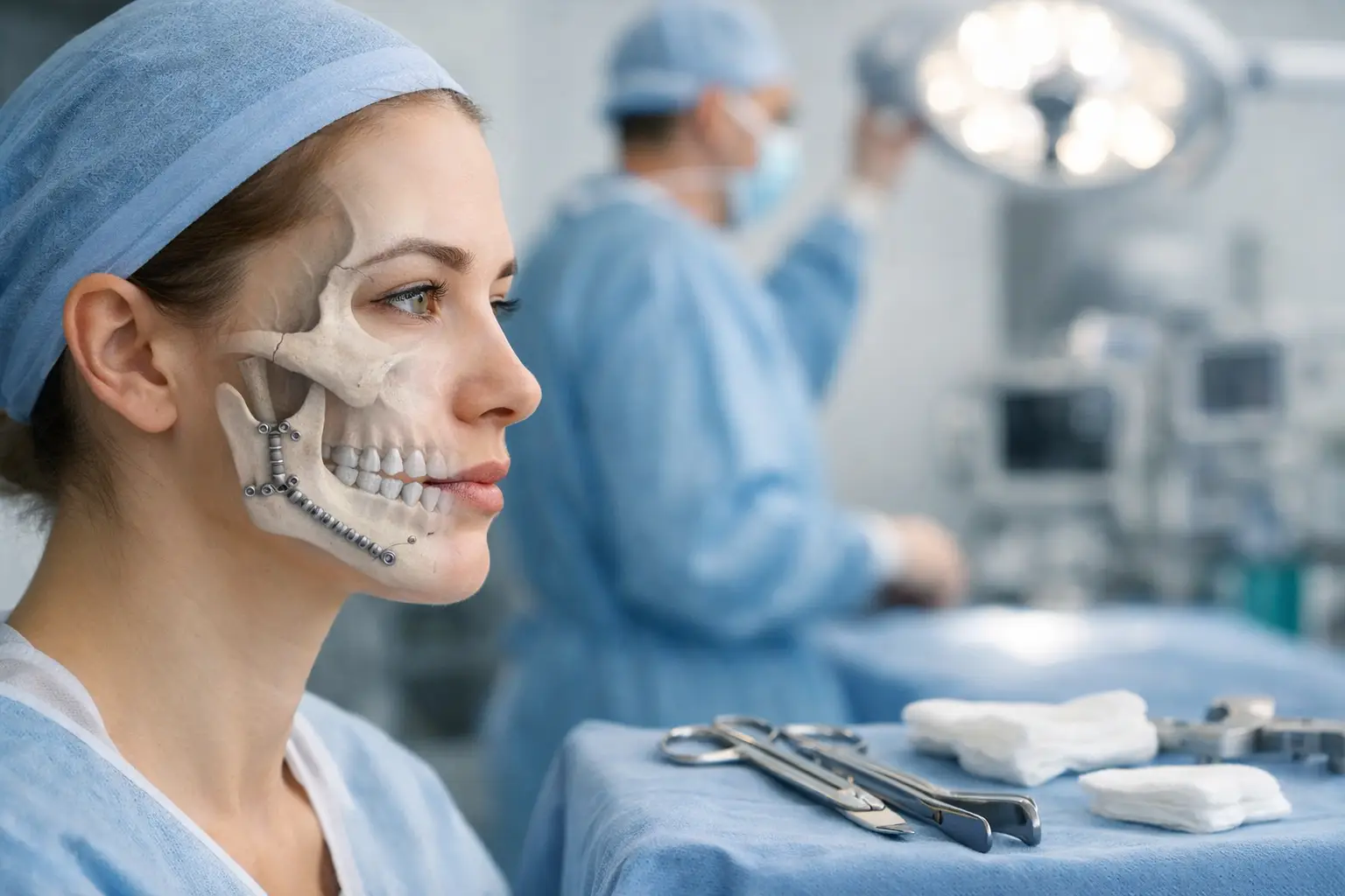
Orthognathic surgery repositions the maxilla and mandible to achieve a balanced occlusal relationship. Le Fort I osteotomy advances or impacts the maxilla in 1-10mm increments. Bilateral sagittal split osteotomy corrects mandibular position through 3-15mm movements. Surgeons use 3D surgical planning software to calculate precise bone movements, ensuring condylar seating and balanced muscle lengths.
Pre-surgical orthodontics decompensates dental positions over 12-18 months. Teeth move into positions that appear worse temporarily but enable optimal surgical correction. Post-surgical orthodontics fine-tunes occlusion over 6-12 months, establishing Class I canine and molar relationships.
Genioplasty addresses chin position when facial balance requires adjustment beyond dental movements. The surgeon cuts the chin bone horizontally, advancing or reducing projection by 4-12mm. Vertical lengthening or shortening corrects facial height discrepancies. The bone segment is fixated with titanium plates, integrating within 8-12 weeks.
⚠️ Important Note
Orthognathic surgery requires 6-8 weeks of modified diet progression – liquids for 2 weeks, soft foods for 4 weeks, then gradual return to normal textures as bone healing permits.
Surgical Planning and Technology
Three-dimensional imaging captures bone architecture at 0.2mm resolution. CBCT scans reveal condylar morphology, airway dimensions, and nerve pathways. Surgeons merge dental models with skeletal data to create virtual surgical simulations. Mock surgeries test different movement combinations to predict soft-tissue changes and the final facial appearance.
Surgical guides transfer virtual plans to the operating room. 3D-printed splints position the jaws precisely during surgery. Cutting guides ensure osteotomy accuracy within 0.5mm tolerance. Custom plates pre-bent to final bone positions reduce operative time and improve stability.
Computer-assisted navigation verifies bone positions intraoperatively. Optical tracking confirms planned movements in real time, enabling immediate corrections. Post-operative imaging validates achieved positions against surgical plans, documenting success within 1mm of intended targets.
What Our Oral and Maxillofacial Surgeon Says
Successful bite correction requires understanding the entire stomatognathic system, not just tooth alignment. Many patients adapt to dysfunction for years before seeking treatment, developing complex compensatory patterns. The surgical approach must address skeletal foundations while preserving or enhancing facial aesthetics.
Timing matters significantly. Younger patients show better adaptive remodelling and faster healing. However, adults benefit from completed growth and stable dental positions. The ideal surgical window balances these factors with the patient’s functional needs and aesthetic goals.
Post-surgical stability depends on achieving balanced muscle forces. Physical therapy restores standard movement patterns while muscles adapt to new jaw positions. This neuromuscular re-education prevents relapse and optimises long-term outcomes.
Putting This Into Practice
- Document bite changes through serial photographs showing tooth wear patterns and facial symmetry from frontal and profile views
- Monitor tooth mobility by gently wiggling each tooth between two fingers, noting any movement or discomfort
- Track TMJ symptoms in a daily log, recording clicking sounds, jaw deviation patterns, and pain locations with activities
- Perform jaw stretching exercises three times daily – open slowly to comfortable maximum, hold 5 seconds, close slowly
- Request a comprehensive bite analysis, including mounted study models and joint imaging, during dental examinations
When to Seek Professional Help
- Persistent jaw pain lasting more than several days
- Visible tooth wear creating flat surfaces or sharp edges
- Tooth sensitivity to temperature changes in multiple teeth
- Difficulty chewing on one or both sides
- Jaw locking in open or closed positions
- Facial asymmetry is becoming more pronounced
- Chronic headaches originating from the temple regions
- Teeth feeling loose or shifting positions
- Clicking or popping sounds with pain during eating
- Limited jaw opening less than three finger widths
Commonly Asked Questions
How long does orthognathic surgery recovery take?
Initial healing requires 6-8 weeks with a modified diet and limited physical activity. Bone consolidation continues for 6-12 months. Most patients return to work within 2-3 weeks for desk jobs, 4-6 weeks for physical occupations. Complete healing with final bite stability occurs at one year.
Can bite problems return after surgical correction?
Proper surgical planning and execution create stable results in most cases. Minor orthodontic relapse may occur without retainer wear. TMJ degeneration or trauma can alter bite relationships. Regular follow-up identifies changes early for intervention.
What determines whether surgery is necessary versus orthodontics alone?
Skeletal discrepancies exceeding orthodontic compensation limits require surgery. Measurements include an ANB angle greater than 7 degrees, vertical facial height discrepancies greater than 5mm, and transverse deficiencies beyond dental tipping capacity. Functional problems like sleep apnea often indicate surgical need.
How do surgeons ensure facial aesthetics during bite correction?
3D planning software predicts soft tissue changes from bone movements. Surgeons balance functional correction with aesthetic proportions using established facial analysis methods. Custom movements address individual aesthetic concerns while maintaining stable occlusal stability.
What happens if uneven bite pressure continues untreated?
Progressive tooth loss occurs through fracture, severe wear, or periodontal destruction. TMJ degeneration leads to chronic pain and limited function. Compensatory patterns create spinal problems. Early intervention prevents irreversible damage that would require more extensive reconstruction.
Next Steps
Uneven bite pressure creates cumulative damage through concentrated forces, TMJ strain, and muscle dysfunction. Early recognition of wear patterns and jaw symptoms enables intervention before irreversible changes occur. Orthognathic surgery provides definitive correction of skeletal discrepancies that cause bite imbalance.
If you’re experiencing persistent jaw pain, visible tooth wear, or facial asymmetry, our Oral and Maxillofacial Surgeon can provide a comprehensive evaluation and treatment options.