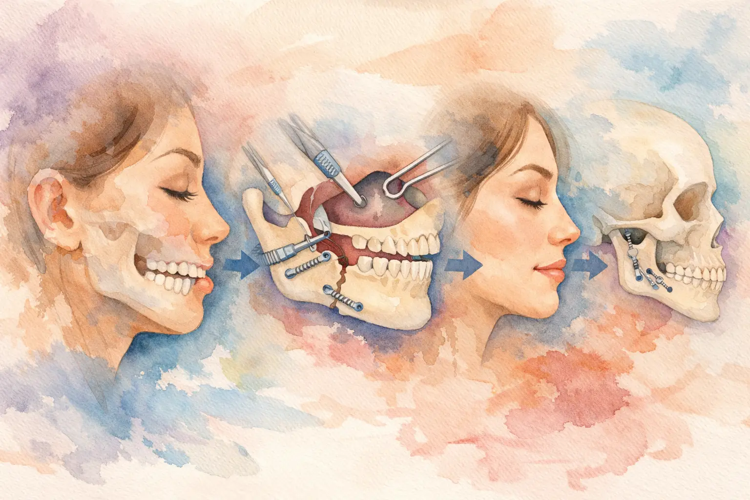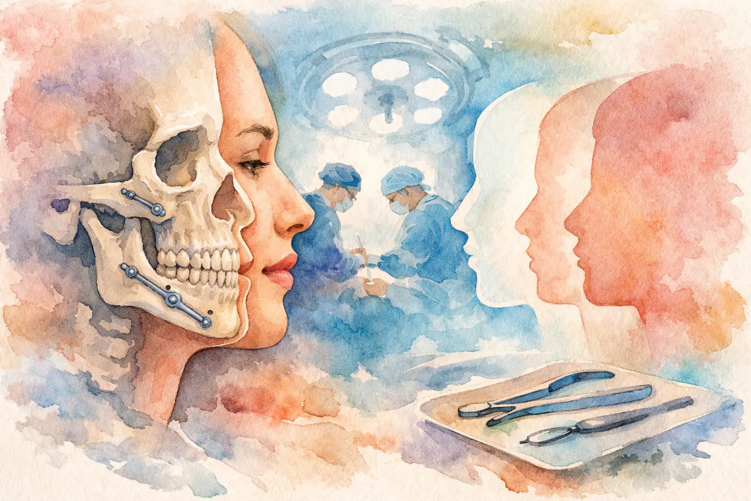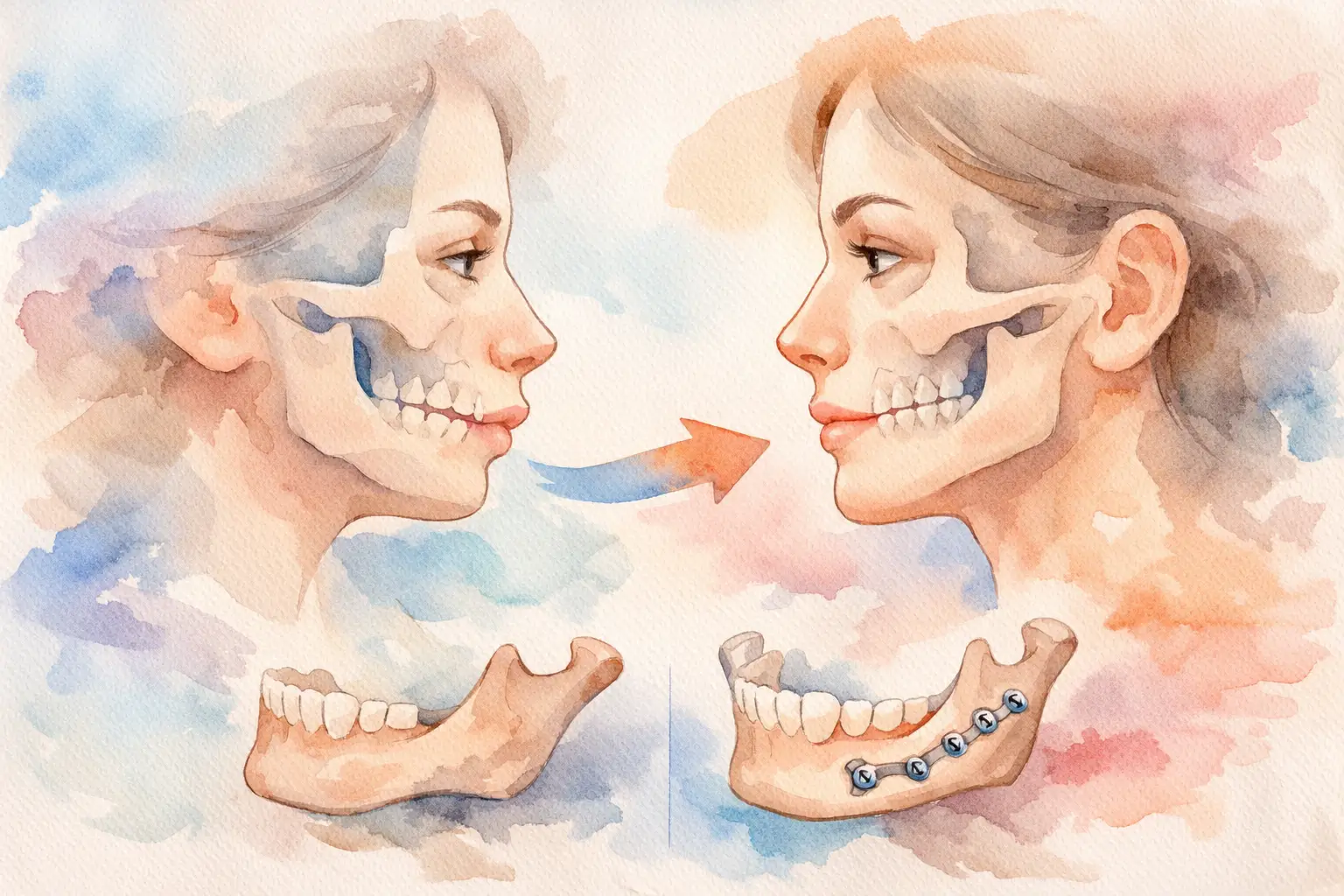
A before-and-after photo can be persuasive in seconds, but jaw surgery before after examples deserve a slower look. A strong result is not just about a more balanced profile or a straighter smile. It is also about how the jaws meet, how the airway functions, whether the lips close comfortably, and whether the treatment plan fits that individual patient.
That is why photo galleries should be viewed as clinical illustrations, not promises. Corrective jaw surgery is highly personalized. Two patients may both have an underbite, for example, yet need very different surgical movements based on facial proportions, dental alignment, airway anatomy, joint health, and treatment goals.
What jaw surgery before after examples can actually show
The most useful examples show more than a cosmetic change. In well-documented orthognathic surgery cases, you can often see improvement in facial harmony, chin position, smile display, and lower face proportion. If the upper jaw was too far back, too long, too short, or canted, surgery may create a more even and proportionate appearance. If the lower jaw was excessively prominent or retrusive, repositioning can improve both profile and bite relationship.
Functional changes matter just as much. Many patients pursue jaw surgery because they struggle with chewing, speech articulation, chronic bite instability, lip incompetence, facial strain, or sleep-disordered breathing. A before-and-after image may hint at those improvements, but the image alone cannot show whether the patient now bites comfortably, breathes better at night, or has less muscle strain in daily life.
This is where context matters. The best examples are paired with an explanation of the diagnosis, the procedure performed, and the treatment objective. Without that, a photo can look impressive while revealing very little about whether the operation solved the actual problem.
Common patterns seen in jaw surgery before after examples
Underbite correction
In patients with an underbite, the lower jaw may appear too far forward, the upper jaw may be underdeveloped, or both issues may be present. Before surgery, the profile can look concave, and the front teeth may not meet properly. After surgery, the facial profile often appears softer and more balanced, while the bite shifts into a more functional relationship.
That said, not every underbite correction looks dramatic in the same way. Some cases are driven mainly by skeletal discrepancy. Others involve dental compensation, asymmetry, or a vertical imbalance that affects the lower third of the face. A good result should look natural, not over-corrected.
Recessed lower jaw
When the lower jaw sits too far back, patients may notice a weak chin-neck transition, difficulty closing the lips without effort, or an imbalanced side profile. In some individuals, a retrusive lower jaw can also contribute to airway narrowing and snoring or obstructive sleep apnea.
After advancement surgery, before-and-after examples often show improved projection of the lower face and a more defined jawline. However, the goal is not simply a stronger profile. The surgeon must also protect bite function, joint stability, and facial proportions from the front and side.
Long face or gummy smile patterns
Some patients show excessive upper gum when smiling or have a vertically elongated lower face. In these cases, upper jaw repositioning may reduce gum display and improve lip competence. Before-and-after examples may show a more balanced smile arc, less strain when the lips are at rest, and a more proportionate lower face.
This type of change is often subtle in isolated photos but significant in real life. Patients commonly report that their face feels more relaxed and that smiling looks more natural rather than forced.
Facial asymmetry
Asymmetry can involve the chin, jaw angles, dental midlines, or the tilt of the upper jaw. These cases are often among the most complex because the issue is not just forward or backward position but three-dimensional imbalance.
After surgery, examples may show improved alignment of the chin with the facial midline, better leveling of the smile, and greater symmetry in the lower face. Still, symmetry in human faces is never absolute. Realistic treatment aims for meaningful improvement, not mathematical perfection.
What photos do not tell you
A polished result photo leaves out much of the treatment journey. It does not show the orthodontic preparation that often comes before surgery, the virtual surgical planning process, the hospital phase, or the swelling that can persist for weeks to months. It also does not show how carefully the surgeon and orthodontist coordinated tooth position with skeletal movement.
Photos also cannot fully capture recovery. Early after surgery, the face may look swollen, stiff, or uneven. Numbness is common in the short term and can take time to improve. Diet changes, activity restrictions, and follow-up care all matter. A responsible surgeon explains these trade-offs clearly rather than focusing only on the final image.
Another limitation is timing. A six-week photo and a one-year photo can look quite different. Early images may still reflect swelling or incomplete soft tissue settling. When reviewing examples, it helps to know when the after photo was taken.
How to assess before-and-after examples critically
First, look for consistency in photography. The head position, facial expression, lighting, and angle should be similar before and after. If the after photo uses better posture, stronger makeup, different lighting, or a more flattering smile, the comparison becomes less reliable.
Second, consider whether the result matches the diagnosis. If the patient had an open bite, do the teeth now appear to close appropriately? If the patient had asymmetry, is the midline improved? If the issue involved airway concerns, was jaw advancement part of a broader functional plan? The photo should support a clinical goal, not just a visual one.
Third, pay attention to whether the outcome looks proportionate. Expert jaw surgery should preserve individual identity while improving harmony. When results look natural, that is often a sign of careful planning. An aggressive change is not automatically a better one.
Why one patient’s result may not predict yours
Even when two patients appear similar, the underlying anatomy may differ significantly. Age, growth status, bone quality, airway dimensions, gum and tooth display, orthodontic needs, TMJ condition, and soft tissue thickness all influence planning and healing. This is one reason why copying someone else’s before-and-after result is not a sound basis for treatment.
There is also the question of expectations. Some patients prioritize bite correction above all else. Others are equally concerned with facial balance, chin position, or breathing improvement. In many cases, the best result is a compromise that respects both function and aesthetics without over-treating either.
This is where specialist evaluation becomes essential. A comprehensive consultation should include facial and dental assessment, imaging, bite analysis, and discussion of goals. For some patients, orthodontics alone may be appropriate. For others, combined orthodontic and surgical treatment offers the most stable and healthy correction.
The role of simulation and planning
Modern jaw surgery is planned with far more precision than a photo gallery can convey. Detailed records, digital imaging, and surgical planning tools allow the team to study jaw position in three dimensions and simulate movements before surgery takes place. That planning helps improve accuracy, but it does not remove the need for judgment. Soft tissue response can vary, and surgery always requires experienced decision-making.
For patients, this means consultation is more valuable than browsing examples for hours. Before-and-after cases can help you understand the range of possible improvements, but your own treatment plan should be based on your anatomy and goals.
When examples are genuinely helpful
Jaw surgery before after examples are most valuable when they help patients ask better questions. What exactly is being corrected? Is the problem in the upper jaw, lower jaw, chin, or several areas together? Will the plan improve chewing, breathing, and facial balance at the same time? What will the recovery period realistically involve?
If the examples lead to a more informed discussion, they have done their job. At a specialist practice such as Aesthetic Reconstructive Jaw Surgery, the real value is not the photo itself. It is the clinical reasoning behind the result, the safety of the process, and the care taken to match treatment to the person rather than the picture.
A useful before-and-after image should make you curious, not certain. The next step is not to compare yourself to someone else’s result, but to find out what your own anatomy is asking for and whether surgery is the right answer.